|
||||||||||
产品简介
产品简介:
| 产品编号 | 产品名称 | 产品包装 | 产品价格 |
| C0005 | 细胞凋亡阳性对照试剂盒 | 200次 | 215.00元 |
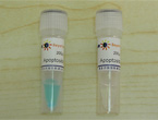
细胞凋亡阳性对照试剂盒(Apoptosis Inducers Kit)含有两种不同的细胞凋亡诱导试剂,试剂A和试剂B。细胞凋亡诱导试剂A(Apoptosis Incucer A,即Apopida)和试剂B(Apoptosis Inducer B,即Apobid)分别可以诱导Hela、HEK293、CHO、COS等常见细胞的细胞凋亡。
由于细胞凋亡的诱导具有细胞特异性,细胞凋亡的机制也具有一定的细胞特异性,尽管细胞凋亡诱导试剂A和试剂B可以诱导常见的一些细胞的细胞凋亡,但是我们无法保证这两种试剂可以诱导任何一种细胞发生凋亡。
细胞凋亡诱导试剂A和试剂B相互补充,可以诱导更多种类的细胞产生凋亡。
本试剂盒至少可用于检测200个样品。
包装清单:
产品编号 |
产品名称 |
包装 |
C0005-1 |
细胞凋亡诱导试剂A |
200μl |
C0005-2 |
细胞凋亡诱导试剂B |
200μl |
― |
说明书 |
1份 |
保存条件:
-20℃保存,一年有效。
注意事项:
对于不同的细胞,需要摸索细胞凋亡诱导试剂的不同浓度和诱导时间。
为了您的安全和健康,请穿实验服并戴一次性手套操作。
使用说明
使用说明:
1. 对于待诱导凋亡的培养细胞,分别按照与细胞培养液的体积比1:1000,1:2000和1:3000的比例把细胞凋
亡诱导试剂A或试剂B加入到培养液中。
2. 4、8、12、16或24小时后观察细胞凋亡的情况。通常16或24小时后,在光学显微镜下可以看到明显的细
胞形态的变化,此时应该可以检测到非常明显的细胞凋亡。
3. 根据初步的实验结果确定一个比较适当的细胞凋亡诱导试剂以及诱导浓度和诱导时间。万一诱导效果不
佳,可以把细胞凋亡诱导试剂A或试剂B同时按照1:1000,1:2000和1:3000的比例加入到细胞培养液中,
以期通过两种不同的试剂同时作用而诱导细胞凋亡。
4. 如果需要在体诱导细胞凋亡,请自行尝试。
产品图片

相关产品
相关论文
使用本产品的文献:
1. Bo J, Yang G, Huo K, Jiang H, Zhang L, Liu D, Huang Y.
microRNA-203 suppresses bladder cancer development by repressing bcl-w expression.
FEBS J. 2011 Mar;278(5):786-92.
2. Zhang JJ, Zheng TT, Cheng FF, Zhang JR, Zhu JJ
Toward the early evaluation of therapeutic effects: an electrochemical platform for
ultrasensitivedetection of apoptotic cells
Anal Chem. 2011 Oct 15;83(20):7902-9. Epub 2011 Sep 20.
3. Xue X, You S, Zhang Q, Wu Y, Zou GZ, Wang PC, Zhao YL, Xu Y, Jia L, Zhang X, Liang XJ
Mitaplatin increases sensitivity of tumor cells to cisplatin by inducing mitochondrial
dysfunction.
Mol Pharm. 2012 Mar 5;9(3):634-44.
4. Wang P, Ren Z, Sun P
Overexpression of the long non-coding RNA MEG3 impairs in vitro glioma cell
proliferation.
J Cell Biochem. 2012 Jun;113(6):1868-74.
5. Yang F, Bi J, Xue X, Zheng L, Zhi K, Hua J, Fang G.
Up-regulated long non-coding RNA H19 contributes to proliferation of gastric cancer
cells.
FEBS J. 2012 Sep;279(17):3159-65. doi: 10.1111/j.1742-4658.2012.08694.x. Epub 2012 Jul
31.
6. Sun DQ, Wang Y, Liu DG.
Cancer cell growth suppression by a 62nt AU-rich RNA from C/EBPβ 3'UTR through
competitive
binding withHuR.
Biochem Biophys Res Commun. 2012 Sep 14;426(1):122-8.
7. Yang F, Sun X, Shen J, Yu LP, Liang JY, Zheng HQ, Wu ZD.
A recombined protein (rSj16) derived from Schistosoma japonicum induces cell cycle
arrest
and apoptosis ofmurine myeloid leukemia cells.
Parasitol Res.2013 Mar;112(3):1261-72.doi:10.1007/s00436-012-3260-8.Epub 2013 Jan 15.
8. Fang XQ, Liu XF, Yao L, Chen CQ, Gu ZD, Ni PH, Zheng XM, Fan QS.
Somatic mutational analysis of FAK in breast cancer: a novel gain-of-function mutation
due to deletion of exon 33.
Biochem Biophys Res Commun. 2014 Jan 10;443(2):363-9. doi: 10.1016/j.bbrc.2013.11.
134. Epub 2013 Dec 19.
9. Zhou Z, Peng L, Wang X, Xiang Y, Tong A.
A new colorimetric strategy for monitoring caspase 3 activity by HRP-mimicking DNAzyme-
peptide conjugates.
Analyst. 2014 Mar 7;139(5):1178-83. doi: 10.1039/c3an02028b. Epub 2014 Jan 21.
10.Qin Y, Chen Y, Wang W, Wang Z, Tang G, Zhang P, He Z, Liu Y, Dai SM, Shen Q.
HMGB1-LPS complex promotes transformation of osteoarthritis synovial fibroblasts to a
rheumatoid arthritis synovial fibroblast-like phenotype.
Cell Death Dis. 2014 Feb 20;5:e1077. doi: 10.1038/cddis.2014.48.
苏ICP备06009238号 |