|
||||||||||
产品简介
产品简介:
| 产品编号 | 产品名称 | 产品包装 | 产品价格 |
| C0011 | 台盼蓝染色细胞存活率检测试剂盒 | 100次 | 96.00元 |
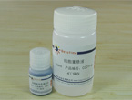
碧云天生产的台盼蓝染色细胞存活率检测试剂盒(Typan Blue Staining Cell Viability Assay Kit),是利用正常的健康细胞能够排斥台盼蓝,而丧失细胞膜完整性的细胞可以被台盼蓝染色研制而成。严格来说,台盼蓝染色检测的是细胞膜的完整性,通常认为细胞膜丧失完整性,即可认为细胞已经死亡。
台盼蓝染色后,通过显微镜下直接计数或显微镜下拍照后计数,就可以对细胞存活率进行比较精确的定量。
台盼蓝染色只需3-5分钟即可完成,并且操作非常简单。
本试剂盒足够检测100个细胞样品。
包装清单:
产品编号 |
产品名称 |
包装 |
C0011-1 |
台盼蓝染色液(2X) |
10ml |
C0011-2 |
细胞重悬液 |
100ml |
— |
说明书 |
1份 |
保存条件:
4℃保存,一年有效。
注意事项:
本试剂盒提供的两种溶液都是无菌的,使用时最好在超净台内进行,避免细菌污染。
台盼蓝对人体有毒,请注意小心防护。
为了您的安全和健康,请穿实验服并戴一次性手套操作。
使用说明
使用说明:
1. 收集细胞:
对于贴壁细胞先用胰酶和/或EDTA消化下细胞。对于悬浮细胞,则可以直接收集细胞。把收集的细胞在
1000-2000g离心1分钟,弃上清,用1毫升或根据细胞的量用适当细胞重悬液重新悬起细胞。
2. 台盼蓝染色:
吸取100微升重悬的细胞到一塑料离心管内,加入100微升台盼蓝染色液(2X),轻轻混匀,染色3分钟
(染色3分钟时间已经足够,但染色时间可以更长一些,但不宜超过10分钟)。
3. 计数:
吸取少量经过染色的细胞,用血细胞计数板计数。通常如果要比较精确地进行定量,每个细胞样品至少
数500个细胞,数出蓝色细胞和细胞总数。
细胞存活率=(细胞总数-蓝色细胞数)/细胞总数X100%
产品图片

相关产品
相关论文
使用本产品的相关论文:
1. Li MX, Wang YR, Cao JM, Tian GZ, Zhao LH.
To Explore the Double - labelingMethod ofMon itor ing the GHRP Regula tory Function on
[ Ca2+ ] i and NO on Rea l Time in Card iomyocytes
Under LSCM.
J Med Res. Aug 2007,Vol.36 No.8.
2. Huang Y, Yang M, Yang H, Zeng Z.
Upregulation of the GRIM-19 gene suppresses invasion and metastasis of human gastric
cancer SGC-7901 cell line.
Exp Cell Res. 2010;316(13):2061-70. Epub 2010 May 15.
3. Kang X, Chen J, Qin Q, Wang F, Wang Y, Lan T, Xu S, Wang F, Xia J, Ekberg H, Qi Z,
Liu Z.
Isatis tinctoria L. combined with co-stimulatory molecules blockade prolongs survival of
cardiac allografts in alloantigen-primed mice.
Transpl Immunol. 2010;23(1-2):34-9. Epub 2010 Mar 22.
4. Chang Y, Yang ST, Liu JH, Dong E, Wang Y, Cao A, Liu Y, Wang H
In vitro toxicity evaluation of graphene oxide on A549 cells.
Toxicol Lett. 2011 Feb 5;200(3):201-10.
5. He XN, Su F, Lou ZZ, Jia WZ, Song YL, Chang HY, Wu YH, Lan J, He XY, Zhang Y
Ipr1 gene mediates RAW 264.7 macrophage cell line resistance to Mycobacterium bovis.
Scand J Immunol. 2011 Nov;74(5):438-44.
6. Wang F, Chen J, Shao W, Kang X, Xu S, Xia J, Dai H, Peng Y, Thorlacius H, Xing J, Qi Z.
The Major Histocompatibility Complex (MHC) of the Secondary Transplant Tissue Donor
Influences the Cross‐Reactivity of Alloreactive Memory Cells
Scand J Immunol. 2011 Mar;73(3):190-7.
7. Peng Y, Zheng Y, Zhang Y, Zhao J, Chang F, Lu T, Zhang R, Li Q, Hu X, Li N.
Different effects of omega-3 fatty acids on the cell cycle in C2C12 myoblast
proliferation.
Mol Cell Biochem. 2012 Aug;367(1-2):165-73.
8. Chen H, Ma N, Xia J, Liu J, Xu Z.
β2-Adrenergic receptor-induced transactivation of epidermal growth factor receptor and
platelet-derived growth factor receptor via Src kinase promotes rat cardiomyocyte
survival.
Cell Biol Int. 2012 Mar 1;36(3):237-44.
9. Ruan J, Song H, Li C, Bao C, Fu H, Wang K, Ni J, Cui D.
DiR-labeled Embryonic Stem Cells for Targeted Imaging of in vivo Gastric Cancer Cells.
Theranostics. 2012;2(6):618-28. doi: 10.7150/thno.4561. Epub 2012 Jun 15.
10.Yi S, Chen Y, Wen L, Yang L, Cui G.
Downregulation of nucleoporin 88 and 214 induced by oridonin may protect OCIM2 acute
erythroleukemia cells from apoptosis through regulation of nucleocytoplasmic transport
of
NF-κB.
Int J Mol Med. 2012 Oct;30(4):877-83. doi: 10.3892/ijmm.2012.1067. Epub 2012 Jul 18.
11.Zeng C, Tang K, He L, Peng W, Ding L, Fang D, Zhang Y.
Effects of glycerol on apoptotic signaling pathways during boar spermatozoa
cryopreservation.
Cryobiology. 2014 Jun;68(3):395-404. doi: 10.1016/j.cryobiol.2014.03.008. Epub 2014
Mar 27.
12.Wu L, Liu YY, Li ZX, Zhao Q, Wang X, Yu Y, Wang YY, Wang YQ, Luo F.
Anti-tumor effects of penfluridol through dysregulation of cholesterol homeostasis.
Asian Pac J Cancer Prev. 2014;15(1):489-94.
13.Su J, Cheng H, Zhang D, Wang M, Xie C, Hu Y, Chang HC, Li Q.
Synergistic effects of 5-fluorouracil and gambogenic acid on A549 cells: activation of
cell death caused by apoptotic and necroptotic mechanisms via the ROS-mitochondria
pathway.
Biol Pharm Bull. 2014;37(8):1259-68.
苏ICP备06009238号 |
